There has been some interaction on our Facebook page referencing Stem Cell Therapy for trigeminal neuralgia. Over the years there has been some controversial claims on the subject, with medical…
Meditation and mindfulness has been shown to not only help with mental health , but also the management of chronic pain. The Federal Government funded mindfulness App Smiling Minds is…
During the webinar presented by Dr Karen McCloy, the issue of how sleep deprivation can severely affect people living with chronic pain was discussed, and she emphasised the importance of…
Transcranial Magnetic Stimulation for Chronic Neuropathic Pain a research trial conducted by the University of California and San Francisco (UCFS) a study on PainPost-Stroke pain Trigeminal Neuralgia Nerve Injury Spinal…
Sometimes people living with trigeminal neuralgia fear that they are invisible, that nobody in the medical field is interested in the disorder and that there is no hope. However there…
The world of artificial intelligence (AI) is likely to enable huge efficiency with remote patient monitoring and interfacing with medical practitioners and research models. This podcast is based on the…
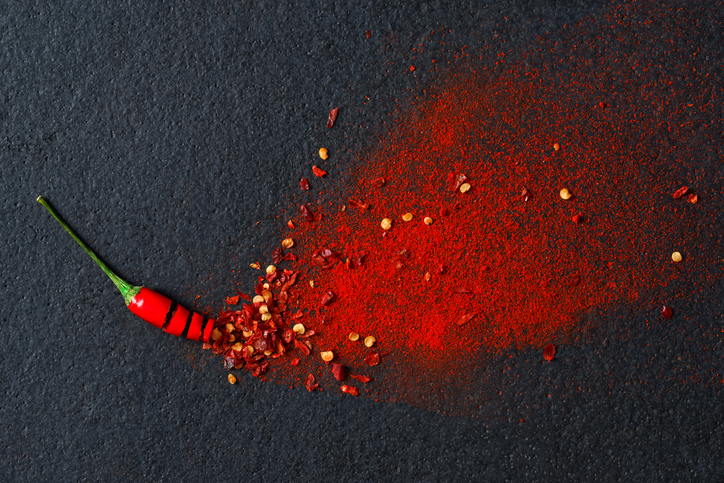
We are constantly looking for treatments which may have an application to relieve neuropathic pain. In 2002 an Australian Doctor Hugh Spencer published his story about utilising capsaicin to cure…
This study evaluates whether Vitamin D supplementation reduces risk of major cardiovascular events in older adults. Vitamin D has also been linked in studies to reduce inflammation and is so…
Research article: Safety of microvascular decompression for elderly patients with trigeminal neuralgia The following is an excerpt of a longer article which can be read in the journal, Clinical Neurology…
STUDY Feasibility of Olive Oil for Reducing Facial Pain of trigeminal neuralgia. Brief Summary: This is a 16-week non-blinded, parallel, controlled trial to determine the feasibility and potential efficacy of…